By Eric Andrew Kristof, RN, CAHIMS | Healthcare IT Professional | HIMSS Arkansas Chapter Member
About This Series: The HIMSS Word of The Day is a running blog series drawing from the HIMSS Dictionary of Health Information and Technology Terms, Acronyms, and Organizations, 6th Edition (2025) — the authoritative reference for healthcare IT professionals and the primary study resource for CAHIMS and CPHIMS certification. Each post paraphrases the term in plain language, grounds it in real-world healthcare IT context, and adds a bedside nursing perspective you won’t find in a textbook.
📖 The Term: Wavelet
Source: HIMSS Dictionary of Health Information and Technology Terms, Acronyms, and Organizations, 6th Edition (2025)
🔍 Plain-Language Definition
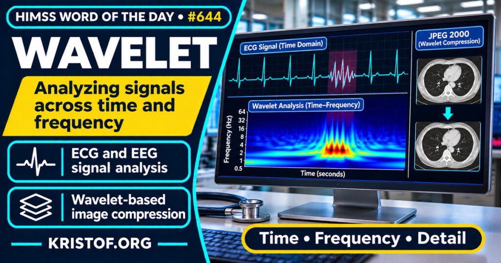
The HIMSS Dictionary describes a wavelet as a mathematical function applied to digital signals and to the shrinking of image files. In the dictionary’s description of wavelet compression, a program analyzes an image, converts it into a set of mathematical expressions, and the receiving end decodes those expressions to rebuild the picture.
The reason wavelets appear in health IT is that clinical signals change over time. Daud and Sudirman (2022) explain that the wavelet approach is favored over the traditional Fourier transform for EEG signals because wavelets are localized in both time and frequency and allow multi-resolution analysis, so brief, transient events can be extracted. Kralj and Lenasi (2022) make a similar point about laser Doppler signals: a classical Fourier analysis cannot localize a signal in time, whereas wavelet analysis provides time and frequency localization. Addison (2005) describes the wavelet transform as a tool for studying complex, nonstationary signals, and calls the ECG a prime example.
| Use | What the sources describe |
|---|---|
| ECG analysis | A review of wavelet transforms applied to the electrocardiogram, using continuous and discrete forms |
| EEG analysis | Reviews of seizure detection and of artifact removal in EEG signals |
| Skin microcirculation | Analysis of laser Doppler signals, with characteristic frequency intervals |
| Image compression | JPEG 2000 is described in imaging studies as a wavelet-based method |
The table is a simplified summary of the reviews and studies cited below.
⚠️ Exam Alert: A wavelet is a mathematical tool, not an image format. JPEG 2000 is a compression method that studies describe as wavelet-based, and JPEG is a related but separate method. The Fourier transform shows which frequencies are present but not when they occur, which is why wavelets suit changing signals. Also keep signal processing, which cleans and analyzes data, separate from compression, which reduces file size.
🌐 Why It Matters: Real-World Healthcare IT Context
Wavelets matter because much of clinical data is a signal or an image. Monitors, waveforms, and imaging systems produce data that has to be cleaned, analyzed, stored, and transmitted, and wavelet methods are one family of tools for those jobs.
What studies show. Addison (2005), in Physiological Measurement, reviewed the role of the wavelet transform in ECG interrogation and described both the continuous and the discrete transform. This is a narrative review from 2005, so newer work is not covered.
Faust and colleagues (2015), in Seizure, reviewed wavelet techniques for computer-aided seizure detection and epilepsy diagnosis, with an emphasis on research from the preceding decade. Daud and Sudirman (2022), in Annals of Biomedical Engineering, reviewed wavelet-based filters for removing artifacts from EEG signals, described the challenges in current work, and recommended alternative solutions. Kralj and Lenasi (2022), in Frontiers in Physiology, reviewed wavelet analysis of laser Doppler microcirculatory signals. They report five characteristic frequency intervals from 0.005 to 2 Hz, each related to a physiological influence, and they note limitations that leave analysis without standardization. These are reviews of research methods, not evidence that wavelet tools improve patient outcomes.
Ko and colleagues (2005), in Radiology, studied wavelet-based JPEG 2000 compression of low-dose chest CT scans on 51 nodules in 23 patients, at ratios of 10:1, 20:1, 30:1, and 40:1. Average measured volume fell as compression increased. There was no significant difference between original and 10:1 images, but there were significant differences at 20:1 and above, and the effect was greater for nodules with ground-glass attenuation. The authors reported a 34.9 mm3 difference for ground-glass nodules at 20:1 versus 8.3 mm3 for higher-attenuation nodules. This is a small study and its findings concern computer-assisted volume measurement.
Penedo and colleagues (2005), in Radiology, evaluated two wavelet-based compression methods, JPEG 2000 and object-based set partitioning in hierarchical trees, on 112 digitized mammographic images from 60 women, at 40:1 and 80:1. Five experienced radiologists read the images, and the authors found no significant difference in detection accuracy between uncompressed images and object-based compression at 80:1. These were digitized film images read by five radiologists in a retrospective study, so the result does not automatically apply to other images or readers.
What I could not find. I did not find a source that states how often hospitals use wavelet-based compression or which vendor products use wavelet filters in monitors. I also did not review work on newer machine-learning methods that combine wavelets with neural networks, so this post does not assess them.
Common wavelet problems:
- Choosing a wavelet type and settings without checking them against the clinical task
- Over-compressing images until measurement accuracy changes
- Assuming a result from one image type applies to another
- Removing real signal while filtering out noise or artifact
- Lack of standard methods, as noted for laser Doppler analysis
- Treating a research method as a validated clinical tool
What supports sound use of wavelets:
- Matching the method to signals that change over time
- Testing compression and filtering with the actual measurement or reading task
- Using modest ratios where measurement precision matters, as the CT study suggests
- Involving clinicians and biomedical engineers in settings and validation
- Documenting which method and parameters were used
- Reviewing the current literature before adopting a new wavelet-based tool
🏥 The Nurse’s Perspective: Clinical, Bedside, & Workflow
Nurses do not choose wavelet settings, but wavelet-based methods may sit behind waveforms and images they rely on.
At the Bedside
Bedside monitors display ECG and other waveforms that are filtered to reduce noise. Whether a particular device uses wavelet-based processing depends on the manufacturer, and that was not verified for this post. What carries over is the practical habit of questioning a tracing that does not match the patient.
In Clinical Workflow
Nurse informaticists often work with biomedical engineering when waveforms or images look wrong, such as a noisy tracing or a blurred image. They can supply the device, time, and patient context so the right team can investigate. The studies above show that the effect of processing choices varies by signal and task.
In EHR Implementation (Go-Live Perspective)
During an Epic Go-Live, device and waveform integration is one part of readiness testing. Analysts at the elbow may hear questions about how a tracing looks in the chart compared with the bedside monitor. This post does not claim that any specific go-live had such a problem. It shows why signal and image quality belong on the test list.
🎓 CAHIMS / CPHIMS & HIMSS Perspective
CAHIMS Exam Domain Mapping
Wavelets connect signal processing, imaging, and data quality:
| CAHIMS Domain | How Wavelet Appears |
|---|---|
| Healthcare & Technology Environments | Biosignals, waveforms, and imaging data in clinical systems |
| Clinical Informatics | Interpreting processed signals and images with appropriate caution |
| Healthcare Information & Systems Management | Compression, storage, and validation of image and signal processing |
| Management & Leadership | Policy for acceptable processing settings and review of new tools |
Key Terms to Know Alongside Wavelet
- JPEG compression — a method for reducing image file size
- DICOM — the standard for storing and exchanging medical images
- Diagnostic Imaging — imaging used to detect and evaluate disease
- Signal processing — methods for cleaning and analyzing signals
- Fourier transform — a method that shows which frequencies are in a signal
- Artifact — a distortion or false feature in a signal or image
HIMSS Organizational Position
HIMSS offers education and research on health information and technology topics through its Resource Center. Members can find related information on the HIMSS Resource Center page.
🧭 More HIMSS Word of The Day Terms
Related terms in the Data and Analytics series:
- Data Visualization
- BTRIS (Biomedical Translational Research Information System)
- Measure
- BioSense Platform
Browse all Data and Analytics terms or the A–Z glossary.
🔗 Explore Further: External References
- Addison (2005), Physiological Measurement — Wavelet transforms and the ECG: a review https://pubmed.ncbi.nlm.nih.gov/16088052/
- Faust et al. (2015), Seizure — Wavelet-based EEG processing for computer-aided seizure detection and epilepsy diagnosis https://pubmed.ncbi.nlm.nih.gov/25799903/
- Daud and Sudirman (2022), Annals of Biomedical Engineering — Wavelet Based Filters for Artifact Elimination in Electroencephalography Signal: A Review https://pubmed.ncbi.nlm.nih.gov/35994164/
- Kralj and Lenasi (2022), Frontiers in Physiology — Wavelet analysis of laser Doppler microcirculatory signals: Current applications and limitations https://pubmed.ncbi.nlm.nih.gov/36741808/
- Ko et al. (2005), Radiology — Effect of CT image compression on computer-assisted lung nodule volume measurement https://pubmed.ncbi.nlm.nih.gov/16126923/
- Penedo et al. (2005), Radiology — Free-response receiver operating characteristic evaluation of lossy JPEG2000 and object-based set partitioning in hierarchical trees compression of digitized mammograms https://pubmed.ncbi.nlm.nih.gov/16244253/
- HIMSS — Resource Center https://www.himss.org/resource-center
Term source: HIMSS Dictionary of Health Information and Technology Terms, Acronyms, and Organizations, 6th Edition (2025). All definitions in this series are paraphrased for editorial purposes. Readers are encouraged to consult the primary source for exact language.
Eric Andrew Kristof, RN, CAHIMS is a Healthcare IT professional and HIMSS Arkansas Chapter member based in Hot Springs Village, AR, with hands-on Epic Go-Live experience and a diverse healthcare and IT background. He writes at kristof.org at the intersection of clinical care and healthcare technology.
Leave a Reply