By Eric Andrew Kristof, RN, CAHIMS | Healthcare IT Professional | HIMSS Arkansas Chapter Member
About This Series: The HIMSS Word of The Day is a running blog series drawing from the HIMSS Dictionary of Health Information and Technology Terms, Acronyms, and Organizations, 6th Edition (2025) — the authoritative reference for healthcare IT professionals and the primary study resource for CAHIMS and CPHIMS certification. Each post paraphrases the term in plain language, grounds it in real-world healthcare IT context, and adds a bedside nursing perspective you won’t find in a textbook.
📖 The Term: Digital Radiography
Source: HIMSS Dictionary of Health Information and Technology Terms, Acronyms, and Organizations, 6th Edition (2025)
🔍 Plain-Language Definition
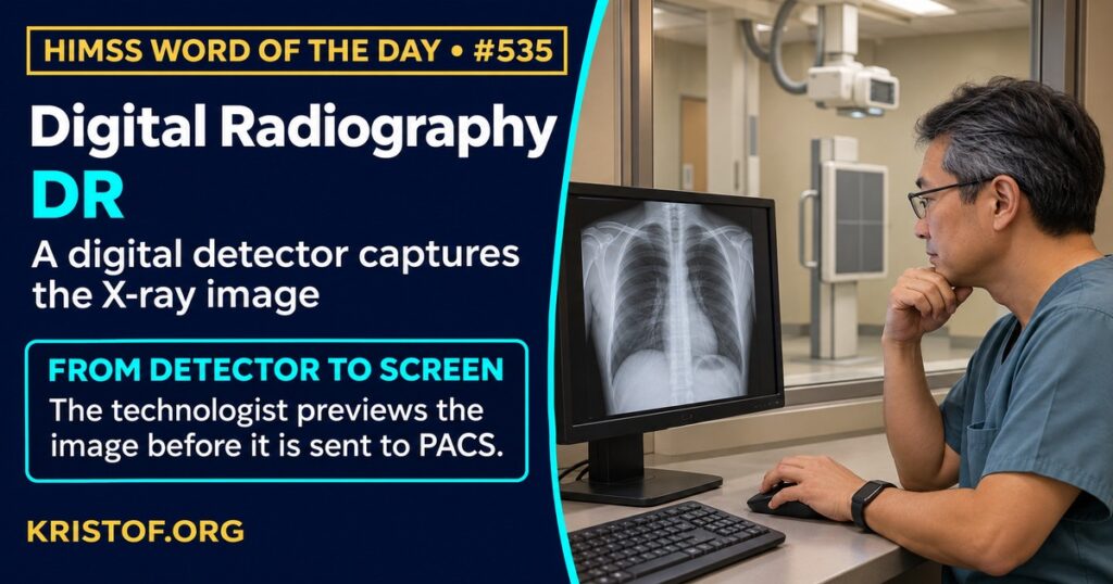
The HIMSS Dictionary defines digital radiography (DR) as radiography that uses a digital x-ray detector to capture images automatically and send them to a computer for viewing. Systems can be fixed or mobile. Because they can handle high volumes, they are commonly chosen for larger or busier clinics and health systems.
The shift from film to digital changed the workflow. In film-based imaging, an exposure produced a physical image that had to be developed. Computed radiography (CR) stored the image on a cassette that was then read in a separate step. Direct digital radiography captures the image in the detector and transmits it electronically, which typically shortens the time from exposure to image and lets the technologist preview the result.
For health IT, digital radiography is both an image source and a data source. Each exam is typically stored in a picture archiving and communication system (PACS), using the DICOM standard, and linked to the order and result in the radiology information system. The detector and software also record technical data: the exposure indicator, body part, projection, and whether the technologist rejected an image and why. Aggregated across units, these data can show variation in radiation dose, image quality, and positioning, and they can support quality assurance. Because images can be retaken quickly, monitoring repeat rates and dose is an important part of quality assurance.
| Item | Role |
|---|---|
| Digital detector | Captures the x-ray image electronically |
| Technologist workstation | Previews, adjusts, accepts, or rejects the image |
| PACS | Stores and distributes images using DICOM |
| Exposure index | Indicates the exposure reaching the detector |
| Reject analysis | Tracks rejected images and their reasons |
⚠️ Exam Alert: Digital radiography (DR) captures the x-ray image directly with a digital detector and sends it to a computer. Computed radiography (CR) uses an imaging plate that is read afterward. Do not confuse DR with PACS (the storage and viewing system), DICOM (the image format and communication standard), or CT, which is a different imaging method.
🌐 Why It Matters: Real-World Healthcare IT Context
Evidence on digital radiography spans systematic reviews, physics studies, and quality programs. Each source below was checked against its published abstract.
Steffensen and colleagues, in Radiography (2021), systematically reviewed acquisition parameters for direct digital radiography. They retrieved 858 studies, screened 89 in full text, and included 23. They found limited evidence to guide changes to accepted best practice, and meta-analysis was not possible because the studies were so different in method. The goals of optimization research varied widely across the studies, and the authors recommend a framework for optimization research.
Scott and colleagues, in the Journal of Applied Clinical Medical Physics (2016), described a quality control program that used exposure index tracking. They studied four digital detectors for a year and compared exposure index histograms across units. They observed differences up to 400 in average exposure index, equal to a 60 percent difference in radiation levels to the detector, between units nominally calibrated to the same exposure index. Distinct peaks reflected the current calibration, a previous calibration, and one that matched computed radiography techniques. The program led to interventions including emphasizing phototimers and improving their calibration. It was a program at one institution, though the authors believe it could be implemented elsewhere.
Tsalafoutas and colleagues, in the Journal of Applied Clinical Medical Physics (2022), used three commercial phantoms and a homemade phantom built to an International Atomic Energy Agency design to examine how exposure parameters affect image quality metrics in a digital radiography unit. The results with the different phantoms were not the same: clear trends of improved image quality with higher air kerma were seen mostly with the IAEA phantom. Variations in scores for repeated identical exposures were almost always below 10 percent for automatic evaluation but reached 17 percent for visual evaluation. The authors suggest post-processing may mask the effect of other parameters. The work was done on one unit with phantoms rather than patients.
Jørgensen and colleagues, in the Archives of Orthopaedic and Trauma Surgery (2023), compared a digital radiography system with a computed radiography system for radiostereometric analysis in 24 hips and shoulders. Mean effective dose for hips was 0.14 mSv with CR and 0.05 mSv with DR, and for shoulders 0.16 and 0.09 mSv. Dose was 64 percent lower with DR for hips and 43 percent lower for shoulders, and there were 60 to 80 percent fewer calibration errors. The authors recommend replacing CR with DR for this use. It is a small, specialized setting, so it should not be taken as a general measure of all exams.
Bantas and colleagues, in the Journal of Medical Radiation Sciences (2023), compared reject analysis between two New Zealand radiology departments using digital detectors over about four months in 2021. They analyzed 47,046 images and 29,279 images and found rejection rates of 7.86 percent and 5.91 percent. Positioning errors accounted for 79.4 percent and 77.3 percent of rejects. Rates and reasons differed significantly by anatomical group, and the authors recommend department-specific interventions. It is a retrospective study of two departments.
Taken together, these sources show that digital detectors can reduce dose in some uses, but that the data they produce, such as exposure indices and reject reasons, must be tracked and acted on to realize the benefits.
Common digital radiography problems:
- Units calibrated to the same target but giving different exposures
- Rejected and repeated images that are not tracked
- Positioning errors as the main cause of rejects
- Post-processing that hides differences in exposure or quality
- Variation in how image quality is scored
- Limited evidence to settle best acquisition parameters
What supports effective digital radiography:
- Exposure index tracking and periodic calibration checks
- Reject analysis by body part, room, and technologist, with feedback
- Phantom testing with a consistent method
- Staff training in positioning and technique
- Integration of detector data with PACS and quality dashboards
- Department-specific targets based on local data
🏥 The Nurse’s Perspective: Clinical, Bedside, & Workflow
Nurses encounter digital radiography at the bedside through portable exams, in the emergency department, and in clinic procedures, so its workflow and safety rules touch nursing practice.
At the Bedside
For portable chest and extremity images, nurses prepare the patient, position lines and devices, and help limit unnecessary repeats. Clear communication with the technologist about patient condition and positioning supports a correct first image. Basic radiation safety practices, such as limiting time and distance, apply to staff as well.
In Clinical Workflow
Nurse informaticists and imaging leaders look at orders, scheduling, and result notifications. Duplicate or unclear orders can lead to repeat exams, and clean order data helps radiology perform the right study. Results that arrive in the record promptly allow nurses to act on findings, such as line placement confirmation.
In EHR Implementation (Go-Live Perspective)
During an Epic Go-Live, order, result, and image links between the EHR and radiology systems get heavy use. Analysts at the elbow hear about missing images, delayed results, and confusing order choices, and they trace them to either build, interface, or workflow causes. Early fixes protect patients from unnecessary delays and repeat exposure.
🎓 CAHIMS / CPHIMS & HIMSS Perspective
CAHIMS Exam Domain Mapping
Digital radiography connects devices, standards, and quality:
| CAHIMS Domain | How Digital Radiography Appears |
|---|---|
| Healthcare & Technology Environments | Imaging modalities, radiation safety, and facility planning |
| Clinical Informatics | Orders, results, image review, and decision support |
| Healthcare Information & Systems Management | PACS, DICOM, interfaces, and detector data management |
| Management & Leadership | Quality assurance, equipment replacement, and staffing |
Key Terms to Know Alongside Digital Radiography
- PACS — system for storing, retrieving, and distributing medical images
- DICOM — standard for medical image formats and communication
- RIS (Radiology Information System) — system that manages radiology orders, scheduling, and reports
- Computed Radiography — x-ray imaging with a reusable plate that is read after exposure
- Exposure Index — indicator of the exposure reaching an image detector
- Quality Assurance — planned activities that monitor and improve performance
HIMSS Organizational Position
HIMSS offers education and research on health information and technology topics through its Resource Center. Members can find related information on the HIMSS Resource Center page.
🧭 More HIMSS Word of The Day Terms
Related terms in the Clinical Informatics series:
Browse all Clinical Informatics terms or the A–Z glossary.
🔗 Explore Further: External References
- Steffensen et al. (2021), Radiography — Optimisation of radiographic acquisition parameters for direct digital radiography: A systematic review https://pubmed.ncbi.nlm.nih.gov/32948453/
- Scott et al. (2016), Journal of applied clinical medical physics — Evaluation of digital radiography practice using exposure index tracking https://pubmed.ncbi.nlm.nih.gov/27929507/
- Tsalafoutas et al. (2022), Journal of applied clinical medical physics — Digital radiography image quality evaluation using various phantoms and software https://pubmed.ncbi.nlm.nih.gov/36345212/
- Jørgensen et al. (2023), Archives of orthopaedic and trauma surgery — Radiostereometric analysis: comparison of radiation dose and precision in digital and computed radiography https://pubmed.ncbi.nlm.nih.gov/36422666/
- Bantas et al. (2023), Journal of medical radiation sciences — Digital radiography reject analysis: A comparison between two radiology departments in New Zealand https://pubmed.ncbi.nlm.nih.gov/36657740/
- HIMSS — Resource Center https://www.himss.org/resource-center
Term source: HIMSS Dictionary of Health Information and Technology Terms, Acronyms, and Organizations, 6th Edition (2025). All definitions in this series are paraphrased for editorial purposes. Readers are encouraged to consult the primary source for exact language.
Eric Andrew Kristof, RN, CAHIMS is a Healthcare IT professional and HIMSS Arkansas Chapter member based in Hot Springs Village, AR, with hands-on Epic Go-Live experience and a diverse healthcare and IT background. He writes at kristof.org at the intersection of clinical care and healthcare technology.
Leave a Reply